Caring for Women
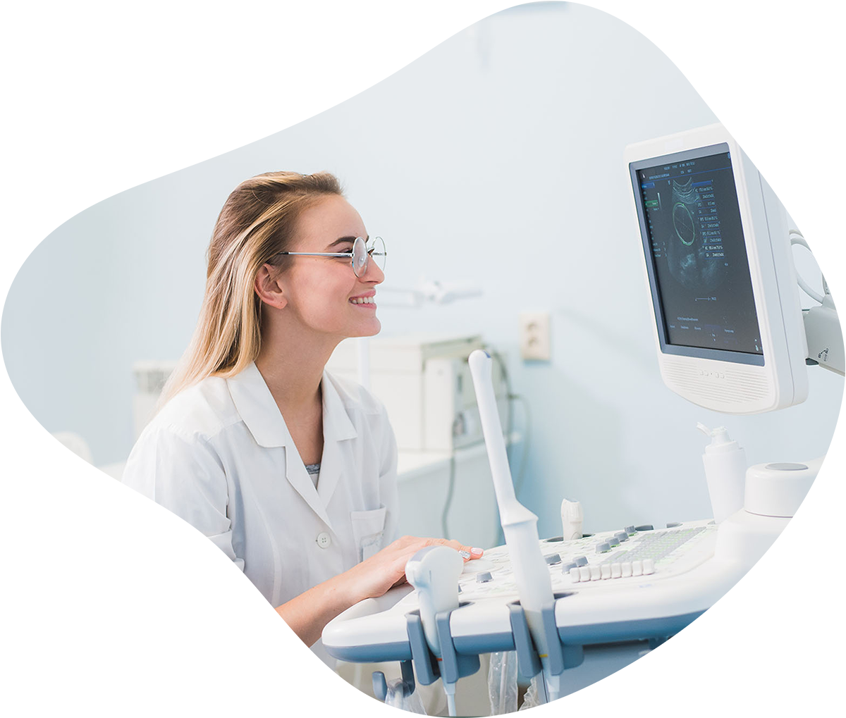
Hawaii Radiologic Associates specializes in providing care for women at our dedicated Women’s Imaging Center in Hilo, and our HRA West Clinic in Kona. The board certified radiologists who interpret these exams are trained in women’s services, including but not limited to breast imaging, fetal ultrasonography and diagnostic evaluation of the female reproductive organs.
We offer a full array of women’s imaging services including 3D screening and diagnostic mammography, ultrasound studies, image-guided biopsies and aspirations and bone density exams (DEXA).
Women’s Imaging Studies
- Digital Screening & Diagnostic Mammogram
- Breast Ultrasound
- Ductography – a mammogram using contrast dye
- Breast Biopsy – through tiny skin incisions using ultrasound or stereotactic mammography guidance.
- Localizations for surgical breast biopsy
- Needle Localization
- DEXA – Bone Density
- OB/Pelvic Ultrasound
- Pelvic MRI
- Hysterosonography – ultrasound of the uterus
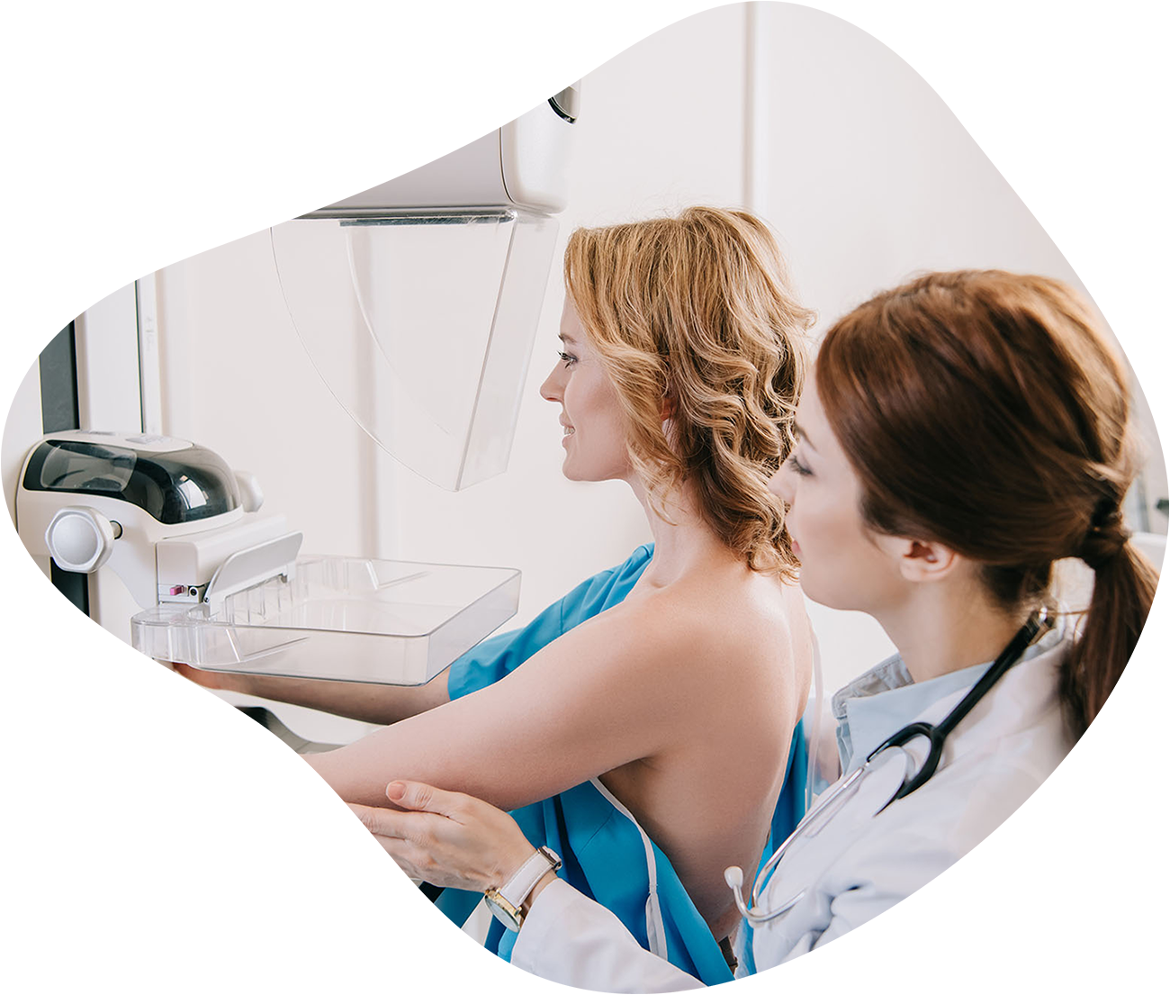
Breast Tomosynthesis
Both of HRA’s Hilo and Kona clinics offer 3D mammography, or digital breast tomosynthesis, which produces multiple X-ray images of the breasts to create a digital 3-dimensional rendering of breast tissue. This allows radiologists to view the breast in 1-millimeter ‘slices’ rather than just the images from the top and side. This technology is particularly helpful for screening women with dense breasts. More than half of Hawaii’s women have dense breasts. Women with dense tissue are four to five times more likely to develop breast cancer.
Our experienced staff of technologists are specially trained to provide the services women need in a skilled, yet reassuring and compassionate manner. HRA provides the finest women’s imaging services in a comfortable environment where the patient’s experience is just as important as the imaging we provide.
Stereotactic Biopsy
When a suspicious breast abnormality is detected by mammography, a Stereotactic Biopsy may be performed. Stereotactic breast biopsy is a safe and minimally invasive form of breast biopsy. It is used to obtain tiny samples from an abnormal breast mass for examination by a pathologist. This biopsy is performed using a computer and x-ray technology to position a hollow biopsy needle. Biopsies are the only definitive way to confirm whether suspicious breast tissue is normal or abnormal.
Ultrasound Guided Biopsy
Following a breast ultrasound, a biopsy using ultrasound, or sound waves, may be performed. Ultrasound imaging is used to assist with proper positioning of the biopsy needle so a small sample of tissue can be obtained and then analyzed under a microscope. Ultrasound guided biopsy is a highly effective way to evaluate abnormal tissue within the breast.
Next Steps:
We’ll report the results
A board certified radiologist will provide your physician with an interpretation of the imaging results.
Your Care Team will report to you
Your physician can then combine these results with your history and explain the findings to you.
We’re Here for You
It’s our specialty to take care of you and your health in times where imaging can be scary. We want everyone to feel welcome and comfortable in every process we provide. Reach out today to get started with our team!